Sunday, January 28, 2007

Rectal polypectomy


Patient who underwent a virtual colonoscopy exam:
a 1.2 cm flat polyp was found in the rectum, top left image
shows the native CT scan axial image, the two images
bottom row are the 3D virtual colon reconstruction,
red arrow: polyp, blue arrow: rectal catheter
Top row: conventional endoscopy, images obtained before
and after polypectomy (red arrow) blue arrow: endoscope.
Images obtained with VOXAR COLONSCREEN software.

Tuesday, January 23, 2007

Rectal Adenoma

A 70-year-old male was referred for a routine CT scan of the abdomen and pelvis with intravenous and oral contrast.
Incidental finding: 4 cm homogeneous nodule high left lateral wall of the rectum.


The patient was referred for colonoscopy and a firm non-bleeding 4 cm mass consistent with adenoma was found 10 cm from the anal margin. The mass was too high to be palpated on rectal exam. Biopsies were obtained. The mass was a villous adenoma, without high-grade dysplasia, it was however too large to be removed endoscopically, and the patient underwent surgical low anterior resection.


It is not uncommon to find large colonic masses on routine scans, especially with thinner cuts obtained with modern multidetector CT scans. We routinely follow the lumen of the colon from rectum to cecum on every single scan (“lumen tracking”). Solid lesions and large adenomas are homogeneous, enhance following contrast injection and can be differentiated from stool in the colon, which usually mix with barium/oral contrast and contain air bubbles.

Rectal mass, endoscopic images

Tuesday, November 21, 2006

Virtual and Conventional Images of a Colon Polyp

Images of a 1.2 cm polyp detected on virtual colonoscopy and conventional colonoscopy images. When a polyp greater than 1 cm is found on virtual colonoscopy it should be removed endoscopically by gastroenterologists.

Colon Cancer Images: Virtual and Conventional Colonoscopy Images

Axial CT scan images of a large sigmoid colon circumferential colon cancer in a 60 year old male, bottom row shows 3D virtual colonoscopy image and corresponding conventional colonoscopy. The patient underwent surgery/sigmoidectomy.

Sunday, November 12, 2006

Preoperative localization of endoscopic findings

We were recently asked by a colorectal surgeon to perform a virtual colonoscopy to localize a 2.5 cm tumor found on endoscopy and described as being " in the distal rectosigmoid". In this case virtual colonoscopy provided useful pre-op evaluation for the surgeon. The coronal cut 3D image and reconstruction simulating double contrast barium enema show the lesion in the distal sigmoid colon (yellow circle). Images obtained with Voxar Colonscreen software.
Click on Images for full screen full resolution

Completion rate of conventional colonoscopy

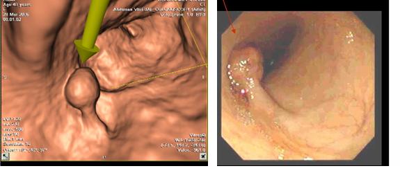
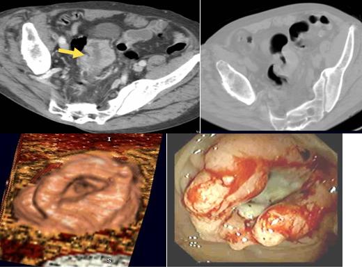
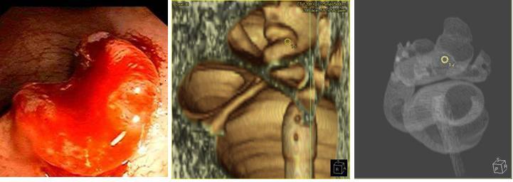
There is some controversy as to what is the exact rate of completion of conventional colonoscopy. Virtual colonoscopy is extremely useful if performed preferably the same day as a failed colonoscopy. The colon is already cleaned, and the proximal colon is usually very well distended on the virtual exam. The exam is much easier to perform than a double contrast barium enema and it has been well established that virtual colonsocopy is more accurate than barium enema.
What is the virtual colonoscopy volume that one may expect from those incomplete colonoscopies?
A recent study published in the New England Journal of Medicine Nov 2006 included 50,148 participants who underwent screening.
The cecal intubation rate/completion rate was 91.1%.
It was somewhat lower than expected for expert colonoscopists. However, the screening colonoscopy program is a large-scale operation that cannot be limited to expert centers (only about one quarter of the 40 centers involved in the study would have been considered expert). Consequently, a less-than-expert rate of intubation probably has to be accepted in a mass-screening setting. Bowel preparation was sufficient, good, or very good for 91.9% of participants.
It is also our experience that in an expert center completion rates approach 96%.
A high volume expert endoscopy center may refer at least 4-5 patients a week for virtual colonoscopy.
Colonoscopy in Colorectal-Cancer Screening for Detection of Advanced NeoplasiaRegula J., Rupinski M., Kraszewska E., Polkowski M., Pachlewski J., Orlowska J., Nowacki M. P., Butruk E. N Engl J Med 2006; 355:1863-1872, Nov 2, 2006

Virtual Colonoscopy Results

New England Journal of Medicine Article
N Engl J Med. 2003 Dec 4;349(23):2191-200. Epub 2003 Dec 1
Pickhardt, M.D et al
Computed tomographic virtual colonoscopy to screen for colorectal neoplasia in asymptomatic adults.
Department of Radiology, National Naval Medical Center, Bethesda, Md, USA. ppickhardt@mail.radiology.wisc.edu
BACKGROUND: We evaluated the performance characteristics of computed tomographic (CT) virtual colonoscopy for the detection of colorectal neoplasia in an average-risk screening population. METHODS: A total of 1233 asymptomatic adults (mean age, 57.8 years) underwent same-day virtual and optical colonoscopy. Radiologists used the three-dimensional endoluminal display for the initial detection of polyps on CT virtual colonoscopy. For the initial examination of each colonic segment, the colonoscopists were unaware of the findings on virtual colonoscopy, which were revealed to them before any subsequent reexamination. The sensitivity and specificity of virtual colonoscopy and the sensitivity of optical colonoscopy were calculated with the use of the findings of the final, unblinded optical colonoscopy as the reference standard. RESULTS: The sensitivity of virtual colonoscopy for adenomatous polyps was 93.8 percent for polyps at least 10 mm in diameter, 93.9 percent for polyps at least 8 mm in diameter, and 88.7 percent for polyps at least 6 mm in diameter. The sensitivity of optical colonoscopy for adenomatous polyps was 87.5 percent, 91.5 percent, and 92.3 percent for the three sizes of polyps, respectively. The specificity of virtual colonoscopy for adenomatous polyps was 96.0 percent for polyps at least 10 mm in diameter, 92.2 percent for polyps at least 8 mm in diameter, and 79.6 percent for polyps at least 6 mm in diameter. Two polyps were malignant; both were detected on virtual colonoscopy, and one of them was missed on optical colonoscopy before the results on virtual colonoscopy were revealed. CONCLUSIONS: CT virtual colonoscopy with the use of a three-dimensional approach is an accurate screening method for the detection of colorectal neoplasia in asymptomatic average-risk adults and compares favorably with optical colonoscopy in terms of the detection of clinically relevant lesions. Copyright 2003 Massachusetts Medical Society